


RETINOBLASTOMA-EYE CANCER
1. What is retinoblastoma?
Retinoblastoma is a cancer of one or both eyes which occurs in young children. There are approximately 350 new diagnosed cases per year in the United States. Retinoblastoma affects one in every 15,000 to 30,000 live babies that are born in the United States. Retinoblastoma affects children of all races and both boys and girls.
The retinoblastoma tumor(s) originate in the retina, the light sensitive layer of the eye which enables the eye to see. When the tumors are present in one eye, it is referred to as unilateral retinoblastoma, and when it occurs in both eyes it is referred to as bilateral retinoblastoma. Most cases (75%) involve only one eye (unilateral); the rest (25%) affect both eyes (bilateral). The majority (90%) of retinoblastoma patients have no family history of the disease; only a small percentage of newly diagnosed patients have other family members with retinoblastoma (10%).
2. Signs and symptoms
Retinoblastoma can present in a variety of ways. The majority of retinoblastoma patients present with a white pupil reflex or leukocoria instead of a normal healthy black pupil or red reflex similar to the one seen when photographs are taken of a child looking directly into the camera. This abnormal white pupillary reflex is sometimes referred to as a cat's eye reflex.
Many times the parent is the first one to notice the cat's eye reflex. Other eye diseases can also present with this white pupillary reflex; leukocoria does not always indicate retinoblastoma. An ophthalmologist can determine the correct diagnosis.
A crossed eye or strabismus is the second most common manner in which retinoblastoma presents. The child's eye may turn out (towards the ear), called exotropia, or turn in (towards the nose), called esotropia.
Retinoblastoma may also present with a red, painful eye, poor vision, inflammation of tissue surrounding the eye, an enlarged or dilated pupil, different colored irides (heterochromia), failure to thrive (trouble eating or drinking), extra fingers or toes, malformed ears, or retardation. On rare occasions, retinoblastoma is discovered on a well-baby examination. Most often, the symptoms of retinoblastoma are first detected by a parent.
When there is a family history of retinoblastoma, newborn babies should be examined in the nursery at birth by an ophthalmologist, or an eye doctor. When there is no family history, it is frequently the parents who notice leukocoria or strabismus and bring their child in for an examination. Often the general ophthalmologist refers the child to an ophthalmologist who specializes in children with retinoblastoma and other cancer of the eye.
The ophthalmic examination by the specialist is best done under general anesthesia. Some very young and older patients can be examined without general anesthesia; this decision is made by the ophthalmologist. When the examination is performed without general anesthesia, the child is placed on his or her back and is wrapped in a sheet like a mummy to restrict the movement of the child's arms and legs. Dilating drops (which sting for approximately 30 seconds after they are placed in the eye) are placed into both eyes prior to the examination. These drops dilate the pupils of the eyes and allow the ophthalmologist to view the retina. Sometimes numbing drops are also placed in both eyes to numb the surface.
If the child is to be examined under anesthesia, the anesthesiologist will put him or her to sleep by placing a mask over his or her mouth or nose. A tube may also be placed in the child's throat to aid breathing and an intravenous line may be started. In order to minimize the risks of anesthesia, the anesthesiologist will ask that the child not be given food or fluids for several hours before the examination. The child will usually fall asleep within a few minutes and the parent may stay until the child is anesthetized. If you have any doubts or questions about whether your child should have anesthesia, you should ask your ophthalmologist, anesthesiologist or nurse.
3. Long term complication of Retinoblastoma.
The majority of children in the United States (over 95%) survive the cancer and have perfectly normal lives. All the children with unilateral retinoblastoma have one normal eye whose sight is not affected even though they may have had one eye removed. Children with one eye have normal vision, play sports, and later drive cars. It is, however, especially important for children with vision in only one eye to wear protective eyewear during sports and other hazardous activities. These children grow up and become famous actors, physicians, lawyers, nurses, accountants, and parents themselves.
The majority of children with bilateral retinoblastoma retain at least one eye with good vision and many are able to retain the use of both eyes. They also live normal lives, go to school, enjoy life, have careers and have families themselves. However, all children with bilateral disease and the 15% of unilateral patients who have the familial form of retinoblastoma will be at much higher risk for other cancers not involving the eyes throughout their lives. Five years after the initial diagnosis of retinoblastoma, more children with the genetic form of retinoblastoma have died from these second tumors than from the original retinoblastoma. The most common second tumors are osteogenic sarcoma, a cancerous tumor which affects the bones, soft tissue sarcomas, and cutaneous melanomas (tumors of the skin, muscle and connective tissue). Although the reported incidences of these tumors vary widely, the risk appears to be about 1% a year. This risk is also increased by the use of external beam radiation, although the amount of increase depends on the age at which the child was treated.
Follow-up appointments are very important when a child is diagnosed with retinoblastoma. All children should be followed by an ophthalmologist and by a pediatric oncologist. Frequency of examinations depends upon the age of the child, the ophthalmologist's suspicion of new tumors, the involvement of one or both eyes, and the type of treatment that the child has received. Mothers and fathers are encouraged to talk to the nurse and to call with questions between visits.
Many parents meet other parents in the waiting room of the physician’s office, clinic, or hospital who also have children with retinoblastoma. Some have found it very helpful to talk to other parents who share similar concerns. Some institutions have newsletters or formal support groups for parents of children with retinoblastoma. Finally, some institutions have programs which can make a child's return to school, home, and the community a bit easier.
Retinoblastoma is a life-threatening disease, but it is rarely a fatal one if treated appropriately. With the correct treatment in the hands of an experienced ophthalmologist and appropriate follow-up both for eyes and for other cancers, the retinoblastoma patient has a very good chance of living a long, full, and happy life.
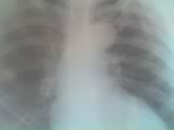
4. Clinical case: a kid of 8 year old Congolese (DRC) living in Goma has leukocoria and strabismus, eye pruritis since 2006. He has been operated 3 times for cataracts and capsular fibrosis as it was diagnosed by doctors in 3 different hospitals but till there the symptoms remain and he continues having the same signs. See pictures in this document. We suspect Retinoblastoma for this kid and we can’t find appropriate treatment for him in the country. He needs support and every one who knows to help him can contact the NGO Agir Ensemble at agirense@yahoo.fr and a referral to St Jude Hospital could be better for him.
5. References
Bramson DH. Pediatric Emergency Casebook: Retinoblastoma. New York: Burroughs-Wellcome, 1985. pp 3-13.
Abramson DH. Retinoblastoma: diagnosis and management. CA: A Cancer Journal for Clinicians 1982. Volume 32, pp 130-142
Abramson DH. The diagnosis of retinoblastoma. Bull NY Acad Med 1988. Volume 64, pp 283-317.
Abramson DH, Ellsworth RM. Ancillary tests for the diagnosis of retinoblastoma. Bull NY Acad Med 1980. Volume 56, pp 221-231.
Abramson DH, Ellsworth RM, Kitchin FD, Tung G. Second monocular tumors in retinoblastoma survivors: are they radiation induced? Ophthalmology 1984. Volume 91, pp 1351-1355.
Abramson DH, Dunkel I, McCormick BM. Neoplasms of the Eye, in Cancer Medicine, 4th ed. Williams & Wilkins, Holland, 1996. pp 1517-1536.
Abramson DH, Servodidio CA. Retinoblastoma in the first year of life. Ophthalmic Paediatric Genetics 1992. Volume 13, pp 191-203.
Char DH, Hedges TR 3rd, Norman D. Retinoblastoma CT diagnosis. Ophthalmology 1984. Volume 91, pp 1347-1350.
Donaldson SS, Egbert PR. Retinoblastoma. In: Pizzo PA, Poplack DG. Principles and Practice of Pediatric Oncology. Philadelphia, PA: Lippincott, 1989. pp555-568.
Dryja, TP. Assessment of risk in hereditary retinoblastoma. In: Albert DA and Jakobiec FA. Principles and Practice of Ophthalmology. Philadelphia, PA: WB Saunders Co. 1996. Volume 5, pp 3270-3279.
Gallie BL, Dunn JM, Hamel PA, et al. How do retinoblastoma tumors form? Eye 1992. Volume 6, pp. 226-231.
Grabowski E, Abramson DH. Retinoblastoma in Clinical Pediatric Oncology, 4th ed. Fernbach DJ and Vietti TJ, ed. 1991. Mosby Books, pp 427-436.
Done in Goma, April 22, 2012.
Mateus Kambale Sahani, M.D.
Agir Ensemble/Goma-DRC.


Aucun commentaire:
Enregistrer un commentaire